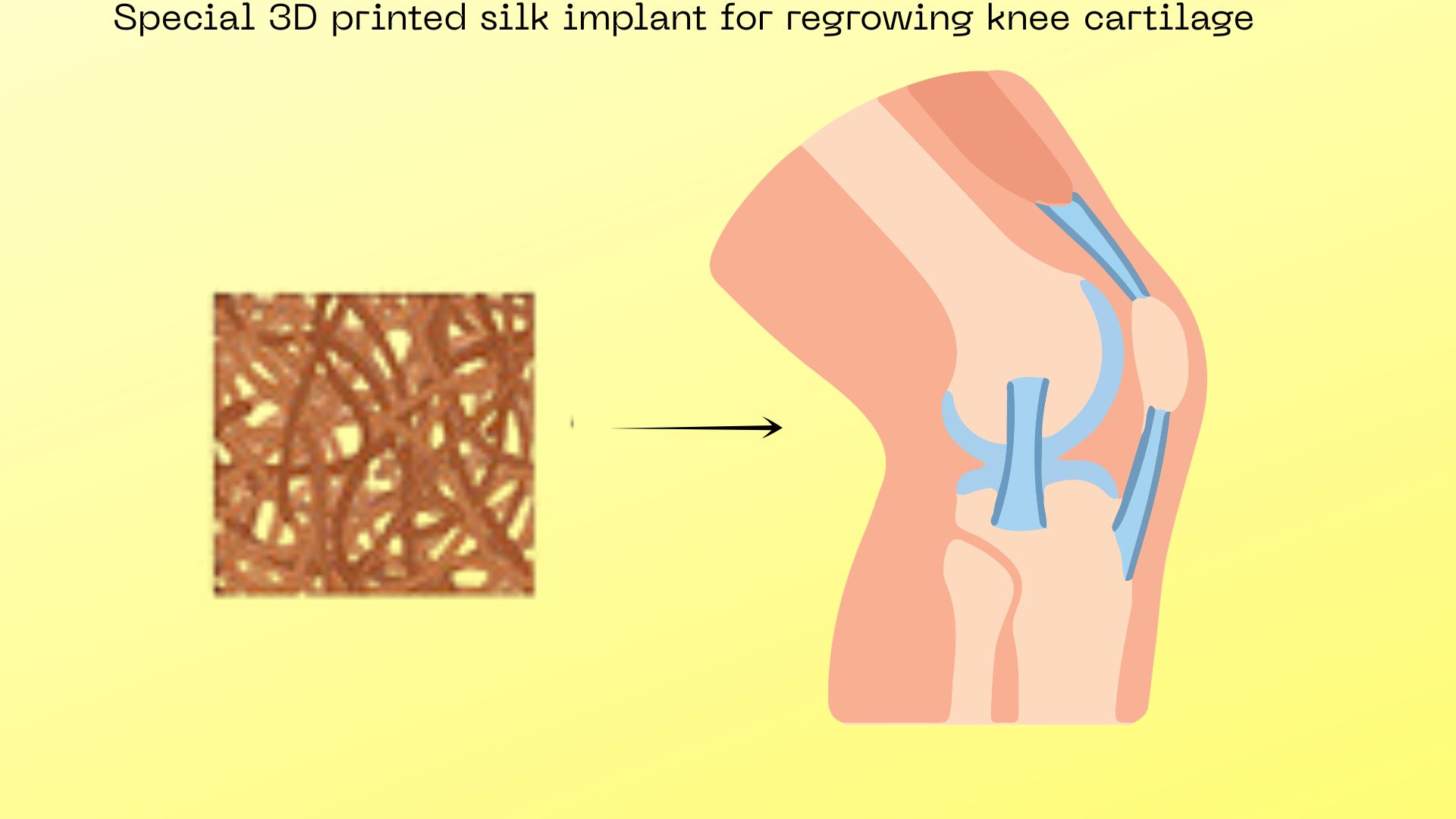
Osteoarthritis affects more than 500 million people worldwide, causing agonizing joint pain as the protective cartilage in the knees gradually wears away. Currently, treatments are limited to managing the pain or waiting until the joint is so damaged that an expensive and invasive total knee replacement surgery is required. Now, an innovative study from researchers at the Indian Institutes of Technology (IIT) Kanpur and IIT Delhi has demonstrated a way to regrow this lost cartilage. Using a novel 3D-printed implant made from silk and gelatin, scientists successfully regenerated healthy, functional knee cartilage in rats suffering from severe osteoarthritis.
When a joint develops osteoarthritis, a hostile, inflammatory environment forms. A specific biological signal known as the bone morphogenetic protein, or BMP pathway, goes into overdrive. This hyperactive signal confuses the cells in the knee, causing them to turn soft cartilage into rigid bone, a process known as hypertrophy. To combat this, the researchers created a biological 3-D printer ink made from silk fibroin, a natural, tough protein derived from silkworms, and gelatin. They then chemically bonded a drug called LDN-193189 directly to the silk. This drug acts to inhibit the harmful BMP signal. At the same time, the silk itself naturally boosts a different cellular signal, known as Wnt, which actively encourages stem cells to grow into healthy, flexible joint tissue called hyaline cartilage.
To test the new method, the research team surgically induced severe osteoarthritis in the knee joints of rats and created a small, critical-sized defect in the remaining cartilage. They then 3D-printed their drug-laced silk constructs to perfectly fit the holes and implanted them into the damaged knees. Over the course of four months, the researchers used micro-CT scans, microscopic imaging, and chemical markers to observe the healing process.
The results showed that in the rats that received the drug-laced silk implant, stem cells migrated from the surrounding bone marrow into the implant, survived the toxic osteoarthritic environment, and transformed into perfectly healthy cartilage. Meanwhile, control groups that received no implant or an implant without the drug saw their joints degrade even further, forming useless scar tissue and painful bone spurs.
While doctors have tried injecting BMP-blocking drugs directly into joints before, the medicines wash away within hours, requiring constant, expensive injections. By covalently binding the drug directly to the 3D-printed silk matrix, this new method ensures a slow, steady release that protects the migrating stem cells long enough for them to permanently establish healthy tissue.
The researchers, however, note that, before treating humans, the implants must first be tested in larger animals, such as dogs or pigs, which more closely simulate the heavy load-bearing conditions of a human joint. The team also hopes to conduct further studies to understand exactly how ageing cells and the immune system interact with the implant to refine the healing Process.
If successfully translated for human applications, this work could change how we treat ageing and joint health. Instead of condemning millions of people to a life of chronic pain, reduced mobility, or major joint replacement surgeries, doctors could one day use this 3D-printed silk implant to coax the body to heal itself. By providing a permanent, regenerative cure for osteoarthritis, this technology has the potential to restore pain-free movement and dramatically improve the quality of life for an ageing global population.