An international team of scientists from institutions in India and the UK has created a new artificial intelligence (AI) tool that serves as a second pair of eyes for doctors diagnosing diseases such as cancer. The system, named HISTO-UNet, analyses microscopic images of human tissue to map out glands and tumours. Unlike previous computer programs, it preserves the exact biological shapes of these structures and uniquely highlights areas where it feels uncertain, ensuring that doctors know exactly where to double-check. The team included researchers from the Indian Institute of Science Education and Research (IISER) Bhopal, Maulana Azad Medical College, Jawaharlal Nehru Cancer Hospital and Research Centre Bhopal, All India Institute of Medical Sciences (AIIMS) Bhopal, and the University of Oxford, UK
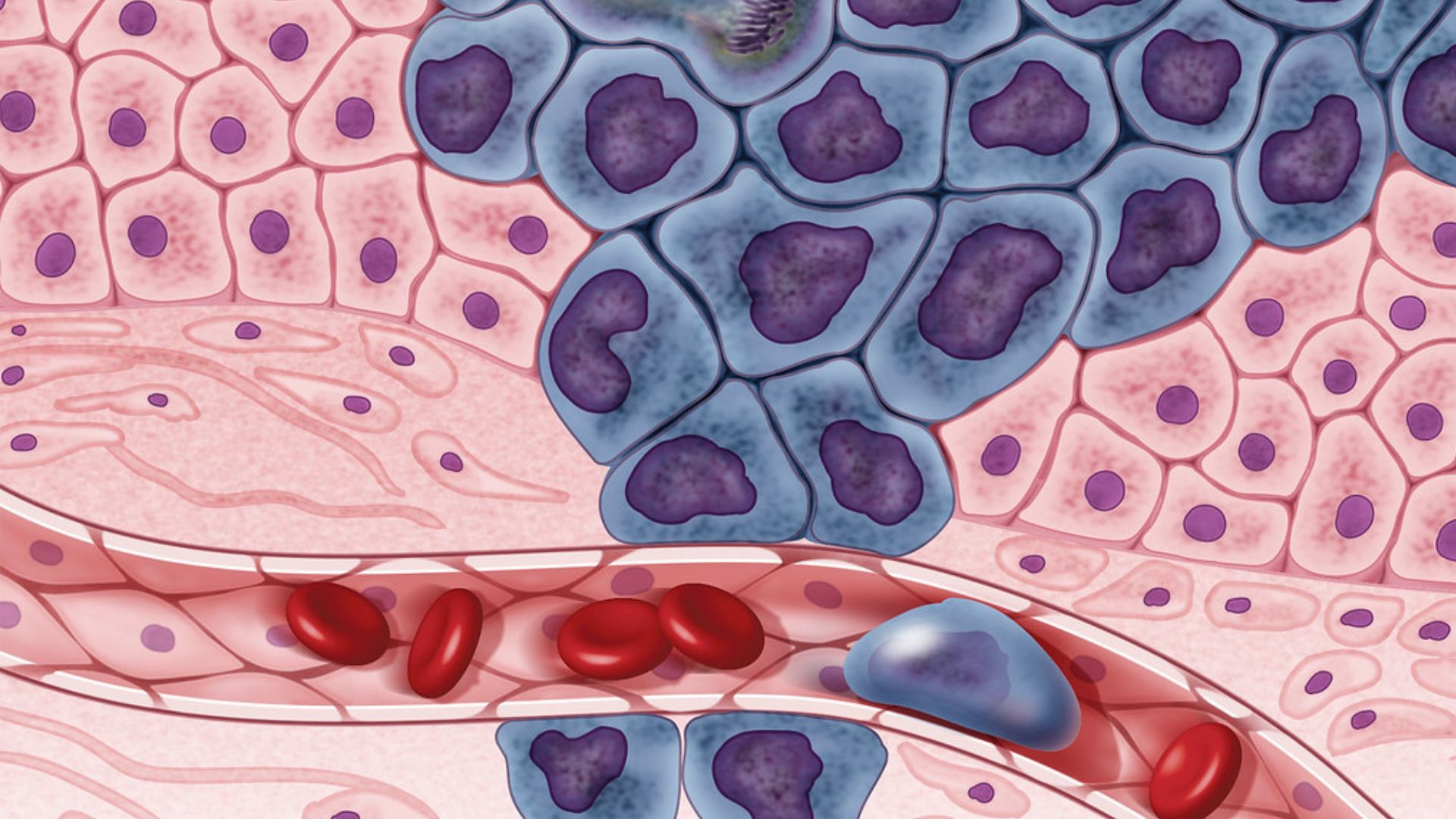
Usually, when doctors examine tissue samples, they are looking for specific shapes that indicate disease. Computational pathology brings the power of computers to the identification of these samples. However, the computational tools were still largely dependent on the medical professionals to identify oddities in the images. Recent advancements in AI, allowing computers to learn from data and improve their functions, is now opening new avenues to medical image identification.
The researchers built HISTO-UNet using a type of AI called a neural network, which mimics human learning. To teach the AI to respect the complex biological shapes of tissues, the team added mathematical rules called topology-preserving constraints. These rules force the computer to recognise the underlying skeleton and the exact centre of a tumour or gland. By doing this, the system is prevented from drawing fragmented shapes, making incorrect connections, or spotting fake holes in the tissue.
Beyond identifying tissue, the researchers also gave the AI the ability to express doubt through a process called dual uncertainty quantification. They programmed the network to run each image through its system twenty-five different times. By looking at how much the AI's answers varied across these attempts, the researchers could measure two specific types of uncertainty. One type measures the inherent fuzziness in the medical image itself, perhaps due to different chemical staining techniques used in the lab, while the other measures the AI's own lack of confidence in its training.
This dual-layered approach marks a significant improvement over previous technologies. Older models, such as the standard UNet algorithm, would simply provide a single guess without any indication of its reliability, which is dangerous in safety-critical medical settings. Furthermore, while some recent experimental models attempted to address shape errors and others to calculate uncertainty, none had successfully combined both tasks into a single, unified system. By bringing these features together, HISTO-UNet consistently outperformed standard models in rigorous testing across three major medical image datasets, demonstrating greater accuracy and reliability. However, the researchers note that because the AI has to process each image multiple times to calculate its uncertainty, it takes slightly longer to produce a final result compared to simpler models.
In busy hospital environments, pathologists often suffer from fatigue while searching through hundreds of complex tissue images. By automatically highlighting the most ambiguous or difficult regions, HISTO-UNet could change the doctor's job from an exhausting, exhaustive search into an efficient review of only the most challenging spots. The system could drastically improve both the speed and safety of disease diagnosis, ultimately leading to faster treatments and better outcomes for patients worldwide.